Key Takeaways
- Research-grade peptide requiring proper handling and storage
- Published studies provide the foundation for ongoing investigation
- Purity verification via HPLC and mass spectrometry is essential
- Mechanism of action involves multiple biological pathways
- Further clinical research is needed to establish translational applications
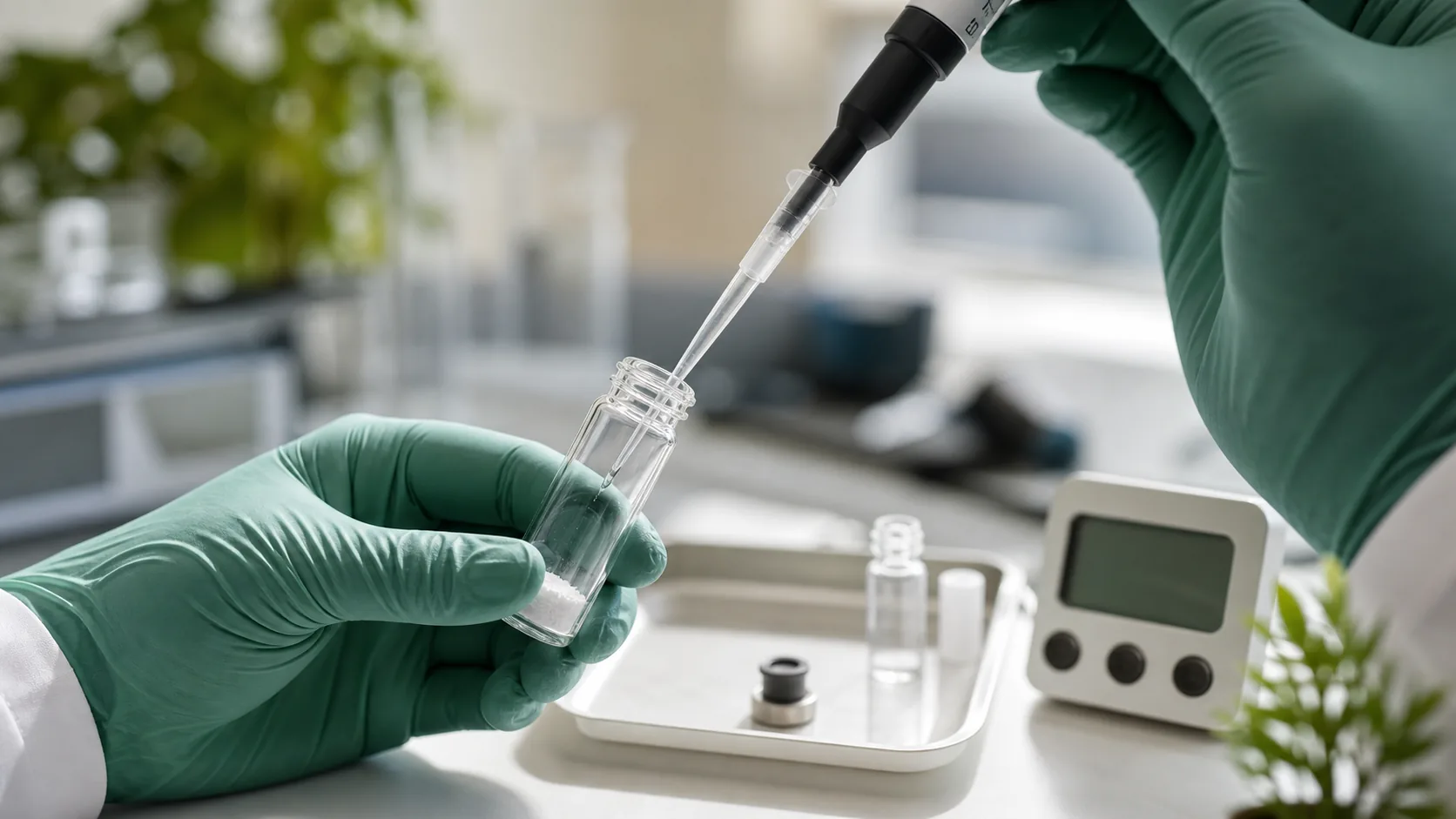
Reconstitution is the critical step between receiving a lyophilized research peptide and preparing it for experimental use. Proper reconstitution technique directly affects peptide integrity, solution concentration accuracy, and experimental reproducibility. This guide covers the science behind reconstitution, diluent selection, step-by-step procedure, and common errors that compromise peptide quality.
Why Peptides Are Supplied Lyophilized
Peptides in solution are susceptible to hydrolysis, oxidation, deamidation, and microbial degradation. Lyophilization (freeze-drying) removes water while preserving the peptide's three-dimensional structure, dramatically extending shelf life. A lyophilized peptide stored at -20°C can remain stable for years, while the same peptide in solution may degrade within weeks.
Diluent Selection Guide
| Diluent | When to Use | Advantages | Limitations |
|---|---|---|---|
| Bacteriostatic water | Most peptides, multi-dose vials | 0.9% benzyl alcohol prevents microbial growth | Not for IV use; benzyl alcohol sensitivity |
| Sterile water | Single-use preparations | No preservatives, purest option | No antimicrobial protection; use within 48-72 hr |
| 0.1M acetic acid | Acidic-soluble peptides (IGF-1 variants, GHK-Cu) | Prevents aggregation at neutral pH | Acidic pH may affect certain assays |
| Normal saline (0.9% NaCl) | Peptides requiring isotonic solution | Physiological osmolality | No preservative; limited shelf life |
| DMSO | Hydrophobic peptides only | Dissolves poorly water-soluble peptides | Cytotoxic at high concentrations; dilute before use |
Step-by-Step Reconstitution Protocol
Materials Needed
- Lyophilized peptide vial with verified Certificate of Analysis
- Appropriate diluent (bacteriostatic water for most applications)
- Sterile syringe (insulin syringe, 29-31 gauge)
- Alcohol prep pads
- Clean, flat work surface
Procedure
- Step 1: Allow both the peptide vial and diluent to reach room temperature (15-20 minutes)
- Step 2: Swab the vial stopper and diluent vial with alcohol prep pads
- Step 3: Draw the desired volume of diluent into the syringe
- Step 4: Insert the needle through the vial stopper at a slight angle
- Step 5: Inject the diluent SLOWLY along the inside wall of the vial. Do NOT spray directly onto the powder
- Step 6: Remove the syringe. Allow the diluent to saturate the powder naturally (1-2 minutes)
- Step 7: Gently swirl (rotate) the vial until the powder is completely dissolved. Do NOT shake or vortex
- Step 8: The solution should be clear and colorless. If cloudy or particulate matter is visible, do not use
- Step 9: Label the vial with: peptide name, concentration, date reconstituted, expiration
- Step 10: Store immediately at 2-8°C (refrigerator)
Common Reconstitution Errors
| Error | Consequence | Prevention |
|---|---|---|
| Spraying water directly onto powder | Incomplete dissolution, local denaturation | Always inject along the vial wall |
| Shaking/vortexing vigorously | Peptide aggregation, denaturation, foam | Swirl gently only |
| Wrong diluent pH | Precipitation, insolubility | Check solubility guide first |
| Excessive volume | Concentration too low for accurate dosing | Calculate volume before reconstitution |
| Contamination | Microbial growth, degradation | Aseptic technique; alcohol swab everything |
| Room temperature storage | Rapid degradation after reconstitution | Refrigerate immediately (2-8°C) |
Peptide-Specific Considerations
| Peptide | Recommended Diluent | Special Notes |
|---|---|---|
| BPC-157 | Bacteriostatic water | Acid-stable; can also use sterile water |
| IGF-1 LR3 | 0.1M acetic acid | Precipitates at neutral pH |
| IGF-1 DES | 0.1M acetic acid | Same as IGF-1 LR3 |
| GHK-Cu | Sterile water | Copper-complexed; dissolves readily |
| Most GH secretagogues | Bacteriostatic water | Standard reconstitution |
Key Research Context
Understanding the research context for How to Reconstitute Peptides: Complete Guide requires consideration of multiple factors including compound purity, experimental design, appropriate controls, and reproducibility standards. The scientific literature provides a foundation for evaluating the biological activity and potential applications of this compound category.
Research-grade compounds require rigorous quality verification before use in any experimental protocol. This includes confirming identity via mass spectrometry, verifying purity via HPLC chromatography (targeting ≥98% for definitive studies), and ensuring proper storage conditions have been maintained throughout the supply chain. A validated Certificate of Analysis from the supplier, ideally with third-party verification, is the minimum standard for quality assurance.
Experimental Design Considerations
Researchers should consider several practical factors when designing experiments with this compound. Dose-response curves should be established using at least three concentration points spanning the expected effective range. Vehicle controls must match the reconstitution buffer exactly. Time-course experiments help determine optimal treatment duration and peak effect windows. For in vivo studies, route of administration significantly affects bioavailability and tissue distribution patterns.
Proper reconstitution technique is essential for accurate dosing. Always inject diluent slowly along the vial wall rather than directly onto the lyophilized cake. Gentle swirling (never vortexing or shaking) prevents aggregation and denaturation. Use bacteriostatic water for multi-dose vials and sterile water for single-use preparations. Record the reconstitution date, concentration, and storage conditions for each vial.
Literature and Evidence Standards
When evaluating the research evidence for any peptide compound, consider the hierarchy of evidence: randomized controlled clinical trials provide the strongest evidence, followed by controlled preclinical studies in validated animal models, then in vitro cell culture studies, and finally computational or theoretical analyses. The number of independent research groups replicating findings, publication in peer-reviewed journals, and consistency of results across different experimental systems all contribute to the overall evidence quality assessment.
Researchers should also be aware of publication bias (positive results are more likely to be published than negative results) and the importance of proper statistical analysis in interpreting study outcomes. Effect sizes, confidence intervals, and appropriate statistical tests are as important as p-values in evaluating research significance. For a comprehensive understanding of peptide quality metrics, review our guide on what 98% purity means and how to interpret analytical data from qualified suppliers.
Methodological Framework
Rigorous research methodology is essential for generating reliable data with any research compound. The following framework outlines best practices for experimental design, quality control, and data interpretation that apply to studies involving this compound category.
Quality Control Protocol
Before initiating any experimental protocol, verify the compound identity and purity through independent analytical testing. The minimum verification standard includes reversed-phase HPLC analysis confirming ≥98% purity and mass spectrometry confirming the correct molecular weight within ±1 Da of the theoretical value. For compounds with disulfide bonds or metal coordination (such as copper peptides), additional analytical methods may be required to confirm proper folding or complexation. Document the lot number, vendor, CoA reference, and storage conditions for every compound used in research.
Dose-Response Characterization
Establishing a complete dose-response curve is fundamental to characterizing any bioactive compound. Use a minimum of five concentration points spanning at least two logarithmic orders of magnitude. Include both sub-threshold and supra-maximal concentrations to define the full response range. Calculate EC50 (half-maximal effective concentration) values using nonlinear regression with appropriate curve-fitting models. For in vivo studies, allometric scaling from published animal data provides initial dose estimates, but species-specific pharmacokinetic differences necessitate empirical dose optimization.
Controls and Replication
Every experiment requires appropriate controls: vehicle controls (matching the reconstitution buffer composition exactly), positive controls (a compound with known activity in the assay system), and negative controls (untreated or inactive analog). Biological replicates (independent experiments on different days with different cell passages or animal cohorts) are more informative than technical replicates (repeated measurements of the same sample). A minimum of three biological replicates is standard for publication-quality data. Statistical analysis should include measures of central tendency, variability (standard deviation or standard error), and appropriate hypothesis testing with correction for multiple comparisons where applicable.
Safety and Handling
All research compounds should be handled according to standard laboratory safety protocols. Wear appropriate personal protective equipment (gloves, lab coat, eye protection) when handling lyophilized powders and reconstituted solutions. Avoid inhalation of lyophilized powder during reconstitution. Dispose of unused compound and contaminated materials according to institutional biosafety and chemical waste guidelines. Research peptides are intended for laboratory research use only and are not approved for human therapeutic use unless specifically noted (such as FDA-approved compounds like Tesamorelin).
Proper storage extends compound viability and ensures consistent experimental results. Lyophilized compounds should be stored at -20°C with desiccant in sealed containers. After reconstitution with bacteriostatic water, store at 2-8°C and use within the validated stability window (typically 3-4 weeks). For long-term storage of reconstituted solutions, prepare single-use aliquots and freeze at -20°C to avoid repeated freeze-thaw cycles that accelerate degradation.
Frequently Asked Questions
How do I reconstitute a peptide?
Inject bacteriostatic water slowly along the vial wall (not onto the powder), allow it to saturate for 1-2 minutes, then gently swirl until dissolved. Never shake. The solution should be clear and colorless.
How long does a reconstituted peptide last?
In bacteriostatic water at 2-8°C: 3-4 weeks. In sterile water without preservative: 48-72 hours. For longer storage, aliquot and freeze at -20°C.
The Bottom Line
This compound represents an active area of peptide research with significant preclinical data supporting further investigation. All research applications require proper analytical verification and adherence to established protocols.
Explore the Research Catalog
All Peptera Research compounds ship with third-party verified Certificates of Analysis.
View CatalogFOR RESEARCH USE ONLY. NOT FOR HUMAN CONSUMPTION. This article is intended for educational and informational purposes only. It does not constitute medical advice. Last updated: April 20, 2026.